
3 March 2023
By: Madisyn Jarvey, Aveya O’Donnell, Gustav Pieper
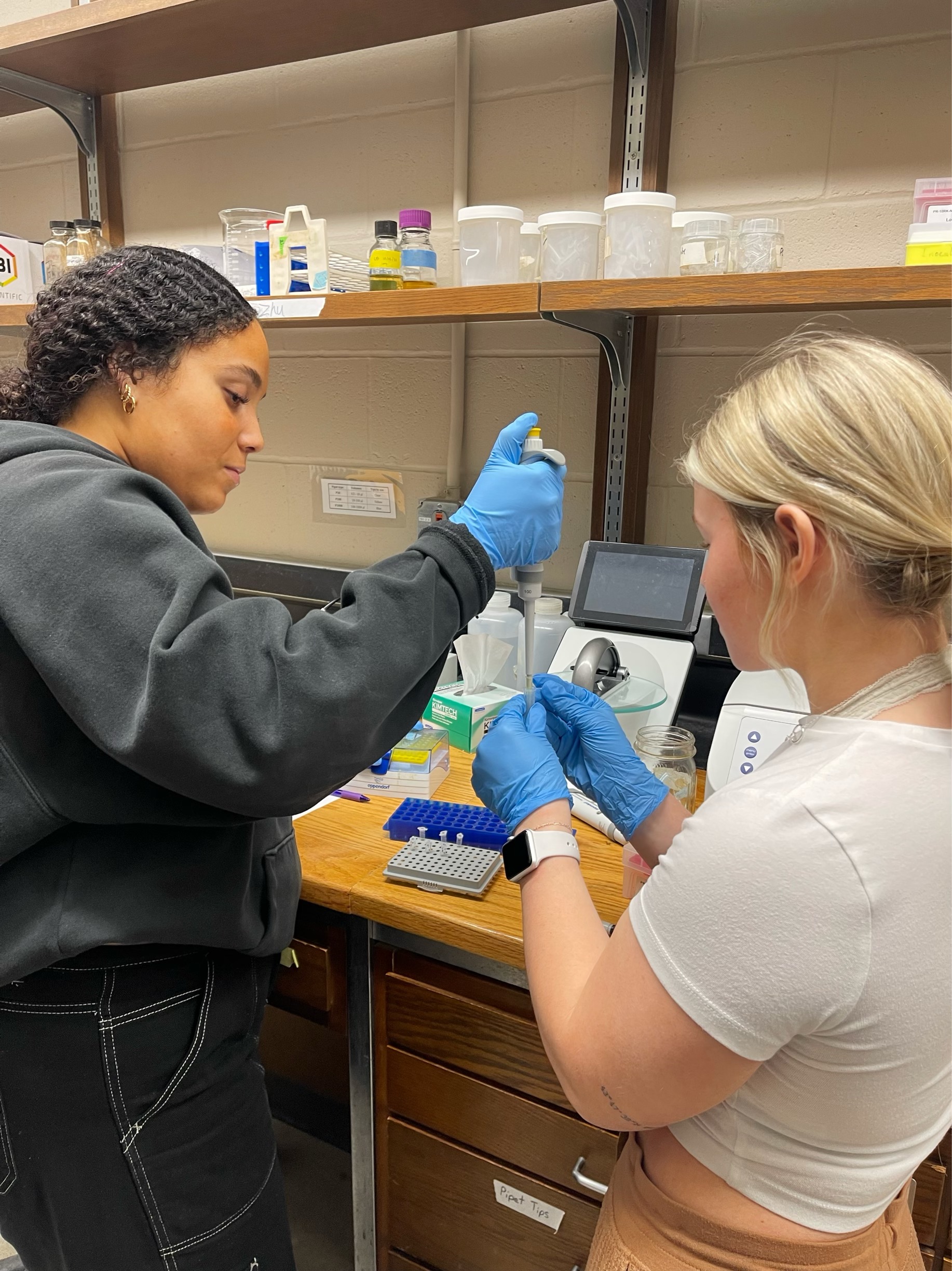
Our group has spent time researching the brain and behavior of green anoles (Anolis carolinensis) lizards. We were instructed to choose a gene and figure out how it affects the brain-behavior of the lizards. The gene we chose to study is Myoneurin (MYNN). Our goal is to figure out if this gene plays a role in the reproductive behaviors of these lizards during the breeding season. Our first step was to design three primers that replicate our MYNN gene. We did this through the National Center for Biotechnology Information website. Once our primers were picked out, we were able to begin the creation of our PCR master mixes, and eventually our gels.
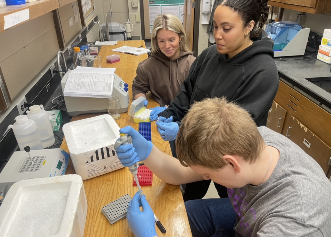
PCR is used to amplify specific DNA sequences. We can observe our primers through gel electrophoresis, which is a technique that will separate DNA fragments by their size. We stain this gel with a DNA-binding dye, and the DNA fragments will be shown as bands. The gel electrophoresis can be made by weighing out 0.75 grams of agarose, which is a powder, and adding 50 ml of 1x TAE to make it a liquid. We then microwave this for around 30 seconds to fully dissolve the agarose. Once the liquid is cooled, we can then place it into the gel casting tray with a comb. The comb creates the wells that we will pipet our PCR into. It takes 30 minutes to solidify so with our downtime, we create new PCR master mixes to run the following week. We did that during our downtime to make sure that we used our time in the lab efficiently.
Over the past couple of weeks, the first PCR that we ran contained the three primer sets that we picked and beta-actin, which consisted of a negative control (H2O) and a positive control (cDNA). Beta-actin is used as a reliable reference gene. For this specific PCR, we used 163 male brain (lizard tissue) as our cDNA. After running our gel and photographing it using a gel imaging bio-doc machine, our results had a lot of issues. Our image revealed primer dimers, incorrect band sizes, and faint bands. For our primers, our band sizes should range between 126-154b. We did not see that in the majority of the PCRs that we ran except for our third primer set. Our first two primer sets were amplifying off large targets. After consulting with Dr. Cohen about our results, we were instructed to run a temperature gradient to see if there are more optimal annealing temperatures. We ran the temperature gradient on our third primer set only because it was the only primer set that gave us decent results throughout the troubleshooting process. We thought that this would reduce our primer dimers and give us clearer results. We run this process through a thermal cycler, which takes the PCR’s through temperatures that denature, anneal, and extend the DNA in the samples.
To be able to start our temperature gradient, we had to multiply our PCR calculations by nine. This was different for us since we were used to multiplying it by two for the master mix. We went through the same process; making gel, pipetting our samples, and putting them in the thermal cycler. After this process was over, we examined the gel and realized that our results were not any different than the other attempts. We still had the incorrect band sizes and primer dimers. Since the temperature gradient did not give us the results we wanted, this tells us that changing the annealing temperatures did not taint our results in the first place. For our primers, we can go back to the normal protocol that we followed before. Our next step was to try a concentration gradient. Our plan for this was to decrease the amount of primer and increase the amount of cDNA. We thought that maybe the bands were faint because of the amount of primer that we had in our PCR rather than the amount of cDNA.
The results of our concentration gradient also did not turn out well. We still had incorrect band sizes and faint bands. We were informed that the bands that we were seeing were just primer dimers and not actual bands. After analyzing the results of our concentration gradient, our future steps will be to rerun our concentration gradient, keeping half of our cDNA samples at 2 μL and the other half at 1 μL but this time, we will be using a different cDNA sample. It could be that our gene is not expressed in this season, and that is why we do not have good amplification of our primers. By using a different cDNA, we will see if that was the issue and improve our results.
In conclusion, this whole process has taught us a lot about patience and perseverance. It is frustrating to not get good results right away, but we will keep working at it.