
April 2, 2021
Emily Schmidtbauer and Kaela Wierenga
Introduction:
In our Bacteria and Disease stream with Dr. Zhu, we are looking at the bacteria Flavobacterium psychrophilum. This bacterium impacts cold water fish and can be seen in the wild but has the biggest impact in fisheries through the spread and containment of the bacteria. We are looking at the gene encoding an endonuclease which catalyzes the cutting of nucleic acids. The purpose of our research is to delete this gene in the bacterium to see what impact is has, if any.
Specific Content:
For our research, we first had to amplify the upstream region of our gene by doing a PCR using a Phusion enzyme and running a gel electrophoresis test. In our first PCR, we used an annealing temperature gradient from 40oC to 60oC to see which would amplify our gene the best. We found that 58.4oC and 47.6oC were the most conclusive. Looking at the other two groups in our stream, we found an average annealing temperature we could use, 52oC. We did not have conclusive bands in our gels, so Dr. Zhu re-ran ours, but at 51oC. This came up with a conclusive band, so we continued with that annealing temperature. We tested our upstream and downstream primers at 51oC and they all had bands appear, but our downstream band was the brightest. Due to this, we decided to continue our research using our downstream primers. We will later look at our upstream primers after some trouble shooting.
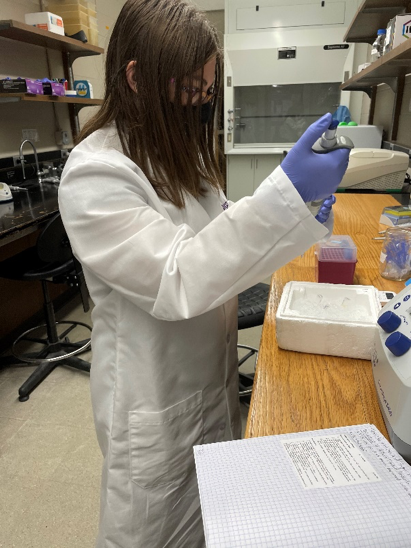
Figure 1. Emily adds LB broth to our E. coli sample.
Our next step was to extract and purify the plasmids, circular DNA, from E. coli samples. We also purified our downstream PCR product to use for future steps. After these were both purified, we checked their concentrations using a NanoDrop spectrophotometer, and they were good enough that we could continue and do a double restriction enzyme digestion. This process cuts at two specific points on the DNA, leaving the circle open, producing two sticky ends. Once this was done, we purified the two samples and ran another gel to make sure we did not lose our DNA in the purification. The bands that appeared on the gel came out faint, but still a good result, so we moved forward to the ligation process. The ligation joined our plasmid open ends with our downstream DNA. To obtain the successively joint products (new plasmids), we did a transformation in E. coli. After mixing the ligation product and E. coli competent cells, incubations on ice, and heat shocks, we added LB broth to our sample. We centrifuged the sample and took out the supernatant to remove any leftover free DNA, leaving the transformed cells in precipitate.
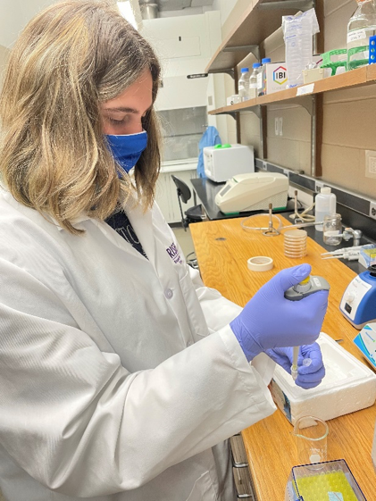
Figure 2. Kaela carefully pipets out the supernatant while leaving the precipitate, E. coli cells, untouched.
Once we had the precipitate, we resuspended the cells in some more LB broth. We put 100 microliters of our sample onto LB agar plates to grow. The plates contained ampicillin and our cells that had the palsmids are ampicillin resistant, so the cells that did not take any palsmids by transformation would not grow colonies. We put the plates in the incubator and will hope for colonies in the next 24 hours. If we get a decent number of colonies, we can continue with our research and do a colony PCR to figure out which colonies have the newly constructed plasmids.
Conclusions/Reflections:
We started with our upstream primers but found that our downstream primers showed better results in gels, so we continued our research with the downstream PCR product. After we do our downstream portion and find results, we will look at doing our upstream region again, but with some troubleshooting. Based on how well our research has been going so far, we believe our future research will go well and give us good results.