
RISE Biochemistry students: Maija Carriveau, Megan Tobias, and Kendall Young
Research Mentor: Dr. Samantha Katner in the Department of Biochemistry, Chemistry, and Geology
Introduction :
The RISE Biochemistry students in the Hepatocellular Carcinoma (HCC) research stream at Minnesota State University Mankato under the supervision of Dr. Samantha Katner have been conducting research since January 2022. This stream allows students to understand sterile practices in the hood such as cell culture and the use of certain drug compounds to target parts of a liver cancer cell. Our goal this semester is to become comfortable working properly with the liver cancer cells so that we will be able to begin more thorough research in the upcoming months.
Research:
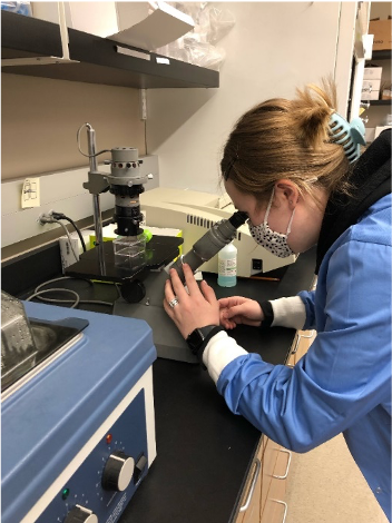
For the 2022 spring semester, we conducted cell culture on 2 types of liver cancer cells in preparation for experiments. First, we set up the sterile hood and got the reagents – trypsin and media – prepared for use on cells. Because trypsin and media are stored in cold temperatures, they need to be warmed up in a water bath to not cause damage to the cells. Once the hood is set up and trypsin and media are warm, we begin to perform cell culture.
The steps of cell culture are done to provide the cells with new nutrients and eliminate dead cells from the flask allowing the live cells to continue growing. Cell culture begins with the removal of the old media carrying with it dead cells. Phosphate Buffer Solution (PBS) is then added to wash the cells. PBS is then removed, and Trypsin is added to detach the healthy adherent cells from the bottom of the flask. After a five-minute incubation period in a 37°C incubator, the flask is removed from the incubator and checked to make sure all the cells are detached. Once proper cell detachment is attained, the flask is placed back into the sterile hood and media is added to deactivate the Trypsin. The collected cells are counted on a hemocytometer and then diluted to a cell concentration for plating in a 24-well plate or in a T-25 flask for future experiments.
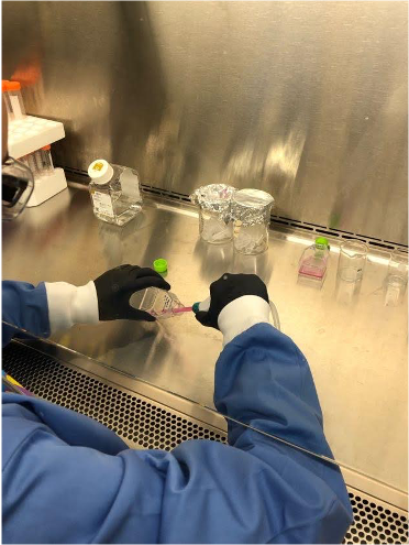
Figure 2: Maija beginning cell culture by taking out old media in flask.

The procedure of cell culture above is used to prepare the cells for assays such as Blyscan assays which we were able to observe and assist in. After the cells were incubated in a 24-well plate, we either collected the cells via trypsin or added concentrations of an enzyme and then collected the cells after the enzyme-incubation. These collected cells were analyzed through a Blyscan assay that measures the number of total proteoglycans present on liver cancer cells. In preforming this assay, we compared the proteoglycans in the two liver cancer cell lines. We counted the cells In addition, we examined cells that have been treated with an enzyme to measure its effects on the proteoglycans.
Conclusion:
Our time in the lab has been a great learning experience, in which we even feel more confident in this lab along with our other general chemistry labs. We have had lots of practice in the tissue culture hood and have found that at first it was hard to get the hang of staying sterile. Now, after making a few initial mistakes that allowed our techniques to improve, we feel confident in our cell culture skills. All the experiments we have performed this semester were fascinating, and when we were able to assist and perform them ourselves that was even more fun! Looking into the future we will be able to perform more assays on the cells to get clearer results on the effectiveness of certain enzymes, and what parts of the cell are being targeted by them. Also, in the future we hope to become comfortable with new lab procedures. We know it will take some time at first but have the confidence to know that we will pick it up in no time. We are excited to continue this research next semester.