
8 April 2022
Jax Karline, Alison Crandell and Corvus Murphy
Introduction:
In our lab, we have just finished confirming the accuracy of our primers in detecting our chosen genes. Our next step is to apply our knowledge about our primers to brain samples collected from Green Anole lizards in various stages of their breeding season. To assess the expression of our genes in the brain, we must look at the levels of our gene’s mRNA found in the brain tissue of these lizards. Since there are limited brain samples, in the past few weeks, our cohort has been practicing honing our newly given skills of RNA isolation with liver samples, which are in more ample stock. When we test genes, we study RNA because DNA examination only provides a static picture of what genes are present. On the other hand, mRNA allows us to see how the gene is being expressed and how much of its corresponding protein is being produced. Unfortunately, RNA samples are volatile compared to DNA, despite the quality and quantity of RNA being critical factors for ensuring the accuracy of gene expression analysis. Therefore, we approached this lab with extra delicacy when performing RNA isolations by extracting the mRNA through a column-based method in our lab.
Research:
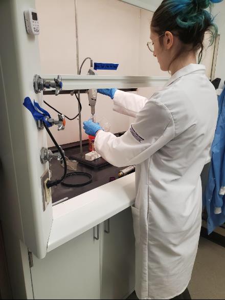
The process of RNA isolation involves homogenizing a tissue sample, separating the mRNA using a centrifuge, and then utilizing a series of columns and buffers to remove the other components of the sample, leaving behind solely the mRNA. We then assess the mRNA sample using a nanodrop to check for concentration and purity and gel electrophoresis to check the mRNA for degradation. This is the most extended protocol we have worked with so far, with time-sensitive steps to prevent the mRNA from degrading. While we were sure to research and review it ahead of time, we still made a few mistakes in our early attempts.
We could not finish our experiment in our first attempt at RNA isolation as we had forgotten to vortex the mixture after adding the chloroform. The purpose of adding chloroform along with phenol is to ensure a clear separation between the aqueous and organic phases. Chloroform and phenol mix well together, unlike phenol and water. Meaning chloroform is necessary for the process as it promotes the separation of the mixture into different layers through the process of being centrifuged. Therefore, we learned the chloroform must be thoroughly mixed with the rest of the mixture before it is centrifuged. Otherwise, the layer with the mRNA does not separate, and it becomes impossible to proceed with the experiment.
Once the chloroform is added and vortexed in, we must centrifuge a sample in a cooled centrifuge, which causes molecules with higher molecular weight to settle down and molecules with lower molecular weight to float. Due to the differing weights, distinct layers will form after centrifuging. In our experiment, mRNA binds to chloroform and maintains a lower molecular weight, making it the first layer in the tube. But an issue that can occur is that if one penetrates the other layers while extracting the uppermost layer, the mRNA sample becomes contaminated and is no longer usable. This means that it is pertinent to be incredibly careful when pipetting throughout this lab, which has proven to be difficult. We have been mostly successful, even though it has caused some difficulty with getting the total volume of the mRNA. Once this topmost layer containing the mRNA is extracted, it is pipetted onto a spin column, allowing fluids to flow through a membrane and into a collection tube. This membrane acts similarly to a filter, and by pipetting different buffers onto the column and centrifuging to flow through, the remaining components of the solution can be washed out and discarded. Eventually, water is pipetted onto the spin column, and after centrifuging, the final flow-through solution will contain only the mRNA.
The results needed to determine if the concentration and purity of our mRNA extractions were in the expected range were the mRNA concentration and 260/280 values. We are expected to produce within the 200-500 ng/µl range for concentrations, but these may increase depending on how many samples may be stuck together. The 260/280 value measures the ultraviolet wavelengths absorbed by the sample and the mRNA has a high quality if it is approximately 2.0. This is a significant value, as an irregular 260/280 value can indicate protein contamination in the sample. We successfully maintained an acceptable ratio for our lab, ranging from 2.08 to 2.14. In addition, we ran our samples through a 1.5% agarose gel to determine mRNA integrity. When performing a gel on mRNA, it is expected to see two clear bands. The gel will not yield distinct bands if the mRNA has broken down. Since our samples did yield the expected bands, it can be concluded that the mRNA had not been degraded during the isolation process.
Reflection:
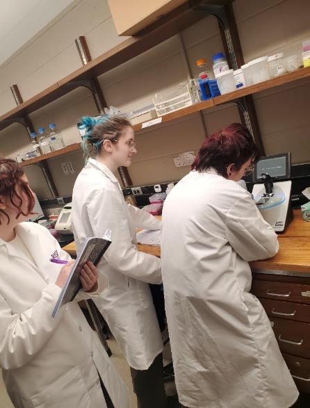
Over the last few weeks, our practice with RNA isolations in the lab has gone smoothly. The few mistakes we made were during our first practices of the newly given protocol, which we quickly caught and were able to recover from. Through these errors, we learned to be extra careful when working in the lab and to ensure that no detail was missed. We carefully read the protocol, underlined the essential information, crossed off each step to keep track of what we have done so far, labeled tubes clearly to keep track of their contents, and worked quickly so as not to let the RNA degrade. Due to these improvements in our lab skills, we have managed to continually produce successful RNA isolations. Now that we have familiarized ourselves with this lengthy protocol and its time constraints, we are ready to begin working with brain samples.