
By D’Aaliyah Johnson & Matthew Trenne
Introduction:
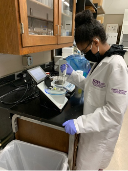
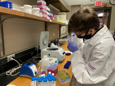
We were selected to be in the “Cancer and Immunity” research stream led by Dr. Land here at the Minnesota State University, Mankato. The primary purpose of this research is to determine how specific MiRNAs interact with proteins to lead to the development of cancer. MiRNA are small fragments of RNA molecules that affect how genes are expressed. We are specifically looking at the genes Hsa-Mir-4297 and Hsa-Mir-12072p. So far in the labs that we have successfully completed we have established a strong control that we will be using as we continue throughout this semester and next.
Specific Content:
So far, we have been experiencing a few bumps in the road regarding our research. Our first lab was DNA isolation, where our goal was to isolate DNA from about one or two million cells. Towards the end of our DNA isolation lab, while we were completing the DNA precipitation, we noticed that the DNA’s pellet was no longer visible once we added 70% ethanol and inverted the tube to wash the pellet. We hoped that it was because the pellet was really clean; therefore, it became translucent, so we continued on with the lab and completed the DNA hydration.
In the next lab, we had to do a PCR amplification. At the beginning of this lab, we nano dropped our clean DNA to determine the concentration. Our ratios were slightly off, making us hypothesize that we had an excess amount of salt in our DNA. We decided to continue on with the lab and create a master mix for our PCR in hopes that the extra salt would not affect our results.
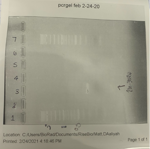
Next, we had to make and load agarose gel for our DNA, whose expected band size is 200-300bp. This is where we experienced the most complications. The first gel that we ran showed great bands for the ladder that we loaded, nothing for the control — which was expected — and nothing for the DNA — which was a problem. We figured that it was because of the excess salt in our DNA, so we followed a protocol to clean the DNA and tried the gel again. Unfortunately, we ran into the same problem with this fresh gel that we experienced with our old gel.
Due to suspected complications with the DNA, we decided to redo the labs, going all the way back to DNA isolation. This time around, we had a lot more DNA when we nano dropped it, so we decided to dilute it. After diluting the DNA, we made another gel and loaded it, and our bands were nonexistent once again. We figured that it was because we did not nanodrop the diluted DNA to see if we had to dilute it some more, so we did that, and the numbers came out perfect.
We decided to make a new master mix using different forward and reverse primers and load a fresh gel. This time we were proud to see the correct size bands of about 200-300bp. As seen in figure 3 well one had our ladder, well 2 was blank, well 3 had our DNA sample, well four was blank, well 5 had the water control, well 6 was blank, and well 7 had another ladder.
Conclusion/Reflection:
We have had a few setbacks in the lab, which is expected while doing research. We believe that those setbacks were due to the primers we have been using for the gels and not the issues with our DNA concentration. This was proven to be true when we switched out our primers with another group and finally saw the expected bands for our 425 DNA on the electrophoresis. We are happy to move on in the research process and hope that we can successfully test our miRNA in the next couple of weeks.