
February 11th, 2022
James Hawco and Jake Lohn
Introduction:
The Bacteria and Disease research stream at Minnesota State University, Mankato, attempts to create a vaccine for bacterial cold-water disease (BCWD) in salmonoid fishes. This process is being done with the deletion of virulence genes in Flavobacterium psychrophilum, the bacterium that causes said disease, to create a weakened version that may be used as a vaccine. Our goal was to delete the fpsY gene in F. psychrophilum using the pYT313 plasmid. The first step is to insert two DNA fragments upstream and downstream of porY into pYT313.
Specific Content:
For the past few weeks, we had conducted plasmid preparation from the Escherichia coli bacterium, otherwise known as E. coli, polymerase chain reaction (PCR), gel electrophoresis, purification, and digestion. We performed PCR with primers to amplify the upstream and downstream regions of fpsY. After PCR, the products underwent gel electrophoresis; this allowed us to visualize and isolate the DNA of interest. The upstream and downstream bands both matched the locations they should be at with the ladder as a reference and were bright to suggest high concentration. Our group compared the products to a 1kb ladder; their product sizes were 2059 b.p for the upstream region and 1912 b.p for the downstream region.
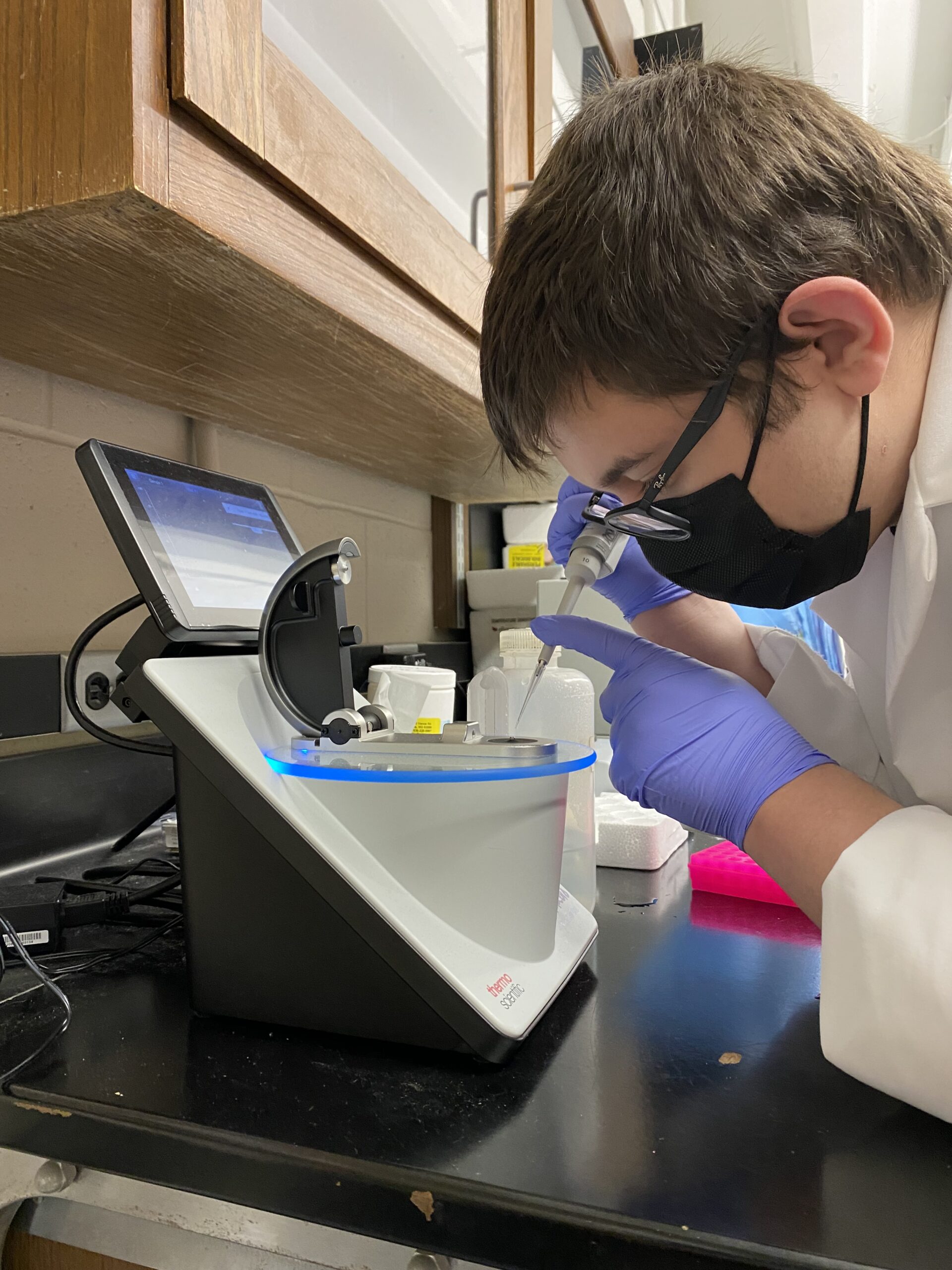
Figure 1: Cleaning the Nanodrop machine before for use to test DNA concentration
We also ran our upstream product through a Nanodrop machine; the results showed that our product had good purity without obvious contaminants. When tested, the results for A260/280 and A260/230 were 1.85 and 2.67, respectively.
Under normal circumstances, the two ratios should be within the range of 2.1~1.8 for A260/280 and above 1.8 for A260/230. Dr. Zhu gave us the thumbs up for our next experiment because the bands that showed up in the gel were single and bright, which suggests a high concentration without nonspecific products. We then conducted double digestion for pYT313 and the upstream region of the target gene using restriction enzymes, creating products that can be used in the next step – ligation.
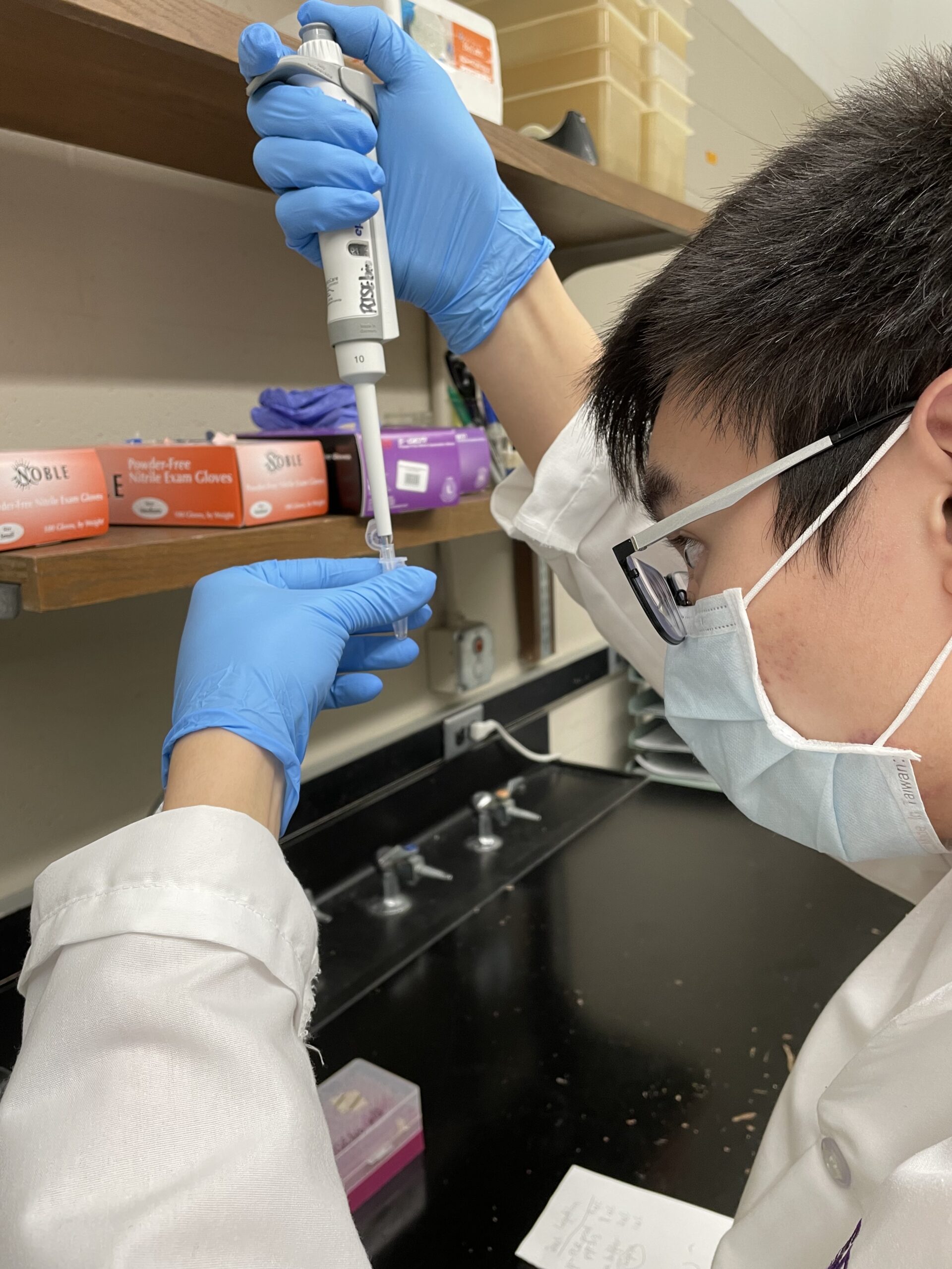
Figure 2: Transferring plasmids from a 1.5ml microcentrifuge tube
Conclusions/Reflections:
Our group has had excellent results so far. We will soon be performing a DNA ligation and inserting our modified plasmids into E. coli to grow colonies of them and perform further testing.